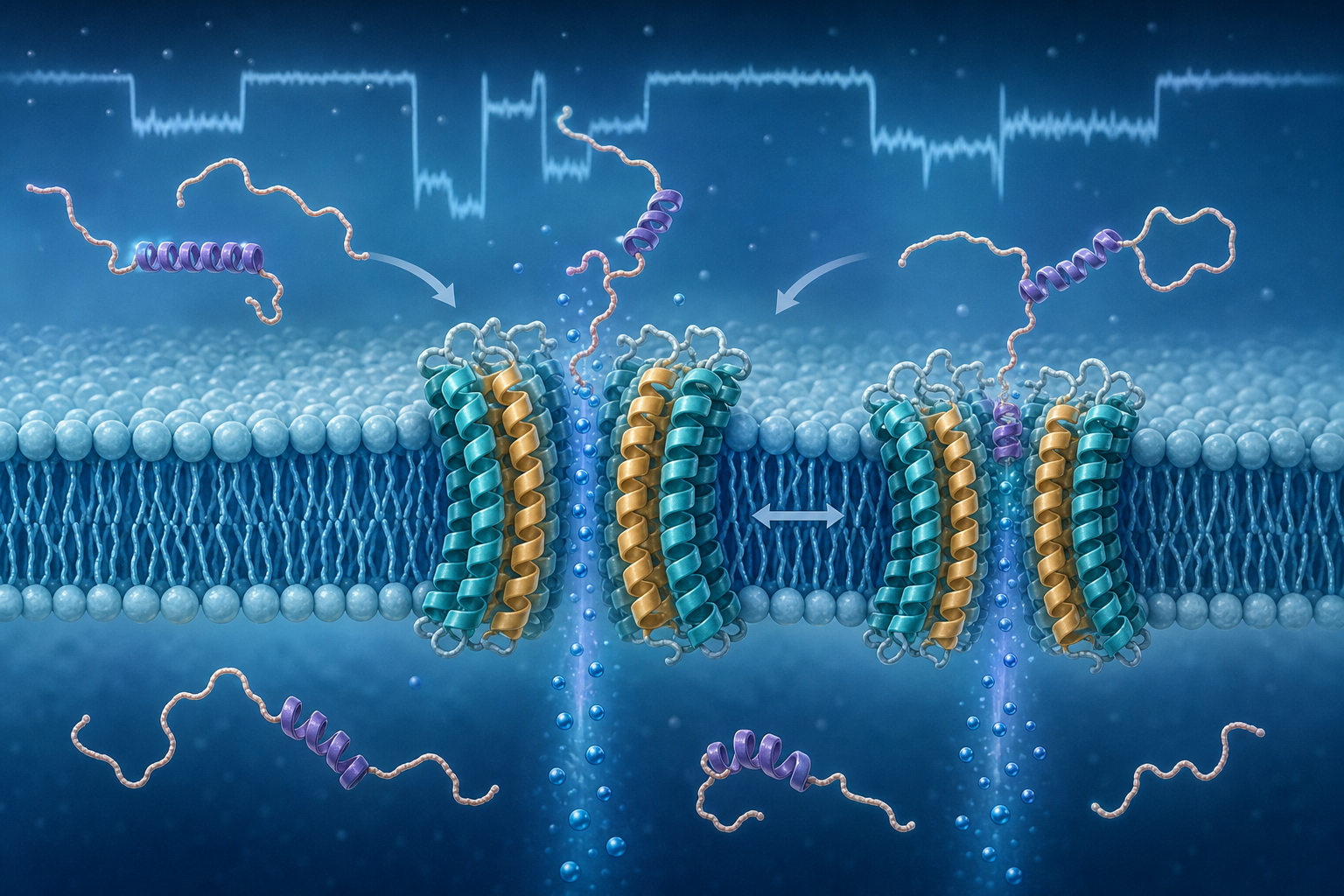
Nanopores measure individual molecules as brief current changes when they pass through or become trapped in a tiny opening. The team developed pPorA, a 40-amino-acid peptide derived from a bacterial porin, which self-assembles in a lipid membrane as an eight-part channel.
The same octamer formed small and large pores with conductances of 2.4 and 3.5 nanosiemens in laboratory electrolyte. Amino-acid and charge changes tuned diameter and selectivity. Experiments were supported by molecular-dynamics models, although their mechanistic explanation uses simplified pores and high electric fields.
Large pores captured several alpha-synuclein variants, including a Parkinson's-associated deletion mutant, with affinity of about 20 nanomolar. Signals distinguished species in mixtures and followed aggregation from monomers through oligomers to fibrils, including changes after an inhibitor. Small pores also detected humanin and SOD1 peptides.
A plausible use is a research chip that rapidly compares how candidate drugs alter toxic protein aggregation, or a complementary biomarker assay requiring very little sample. The study did not demonstrate diagnosis from patient blood, clinical accuracy or the ability to identify disease from one signal.
Translation requires reproducible pore manufacture, automated signal interpretation, testing in blood or cerebrospinal fluid, large blinded patient cohorts and comparison with established methods. Optimistically, specialist research chips could emerge in 3–5 years. A diagnostic test, if validation succeeds, is unlikely before 7–12 years, and broad use remains uncertain.
Be the first to open the discussion.