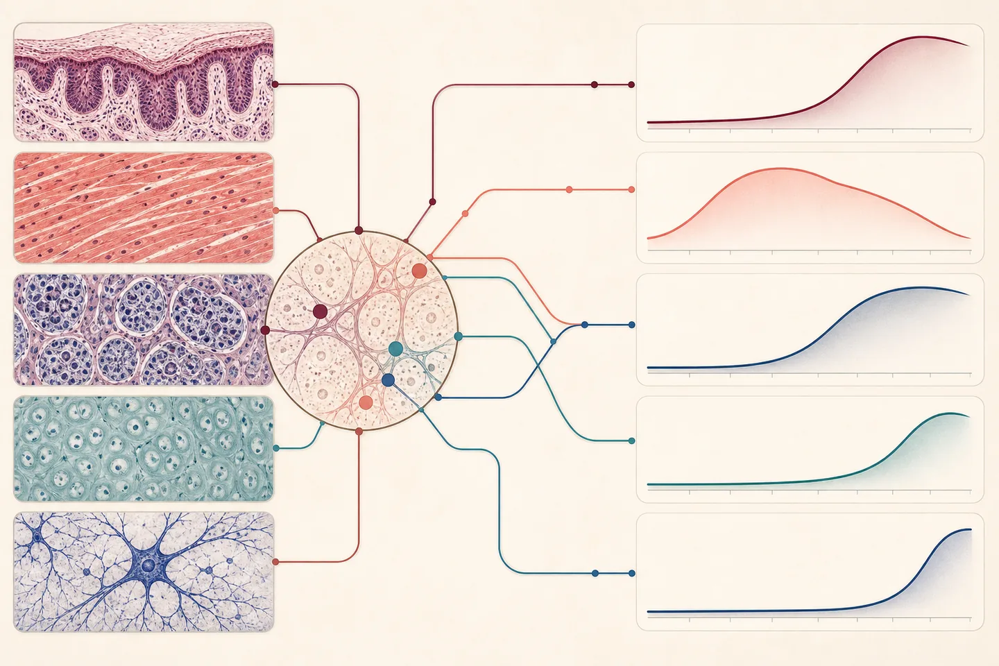
Aging is often reduced to one biological age, yet the architecture of individual tissues changes differently. The authors developed PathStAR, which uses a pretrained digital-pathology model to convert tissue images into visual features and compares adjacent ten-year age windows.
They applied it to 25,306 post-mortem biopsies from 40 tissues in 970 GTEx donors aged 21–70. Fifteen tissues passed the filters for detailed trajectories. Vascular tissues changed most rapidly around the thirties, uterus and vagina mainly around menopause, while most examined digestive and male reproductive tissues showed two peaks of change.
Across organs, faster structural change coincided with stronger inflammatory pathways and reduced energy production, repair and cellular quality control. Deviations in some related organ systems were also coordinated within the same donor. These are associations, however, not proof that one identified mechanism caused the changes.
If independently confirmed in longitudinal cohorts, the map could help decide when a particular organ should be monitored or a preventive intervention tested. PathStAR might also tell researchers whether a candidate treatment changes the microscopic structure of the intended tissue instead of relying on a single whole-body aging marker.
Translation requires validation in living patients, repeated samples over time, broader ages and populations, standardized slide preparation and evidence that the score predicts health better than conventional risk measures. Optimistically, a retrospective research tool could mature in 2–4 years and limited clinical validation begin in 5–8 years. Routine testing is more plausibly 8–15 years away, if it proves useful at all.



Be the first to open the discussion.