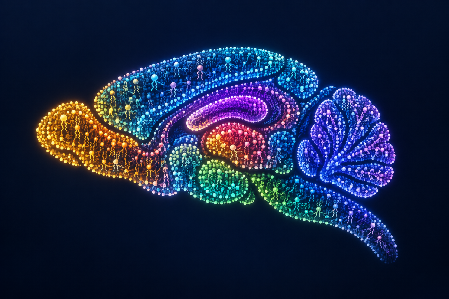
Lipids are more than passive building material in the brain. They form cell membranes and the myelin around nerve fibres and help signals travel. In a peer-reviewed Nature study published on 23 September, an EPFL team mapped 172 lipids across 109 sections from 11 mouse brains. It assembled a three-dimensional atlas from about 7.2 million measurement points.
The analysis divided the tissue into 539 territories defined by similar lipid mixtures, which the authors call lipizones. Some align with familiar anatomical regions; others expose differences within white matter or connect nerve-cell bodies with distant endings of their fibres. The atlas adds a chemical layer to cell and gene-activity maps. The number of territories does not mean 539 previously unknown brain parts were discovered.
In three pregnant mice, the team observed shifts in lipid patterns across several regions, notably the cortex and white matter. That small group and single studied condition cannot establish how a human brain changes during pregnancy or what causes the shifts. The study supplies neither a treatment nor a test for neurological disease.
The public atlas could help researchers decide which lipid patterns to investigate in development, ageing and disease. Independent replication, other species and direct links between chemical maps and cell function are still needed. Follow-up comparative studies might optimistically appear within two to five years; there is no sound timetable for human use.


Be the first to open the discussion.