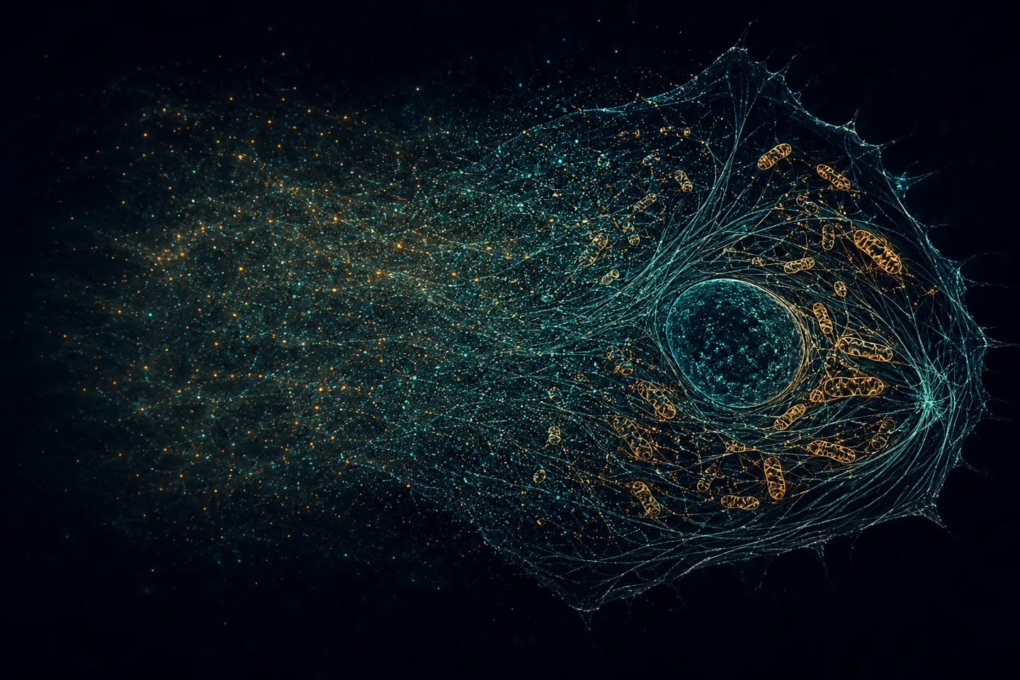
Super-resolution microscopy usually relies on intense laser excitation, which can bleach labels and harm living cells. A Nature team has introduced RIED, a computational method that reconstructs sharper images from irregular photons emitted by electrochemiluminescence, chemiluminescence or bioluminescence, without external laser excitation.
In fixed COS-7 cells the authors report lateral mitochondrial resolution of 102 nanometres and axial resolution of 221 nanometres; bioluminescence in living cells reached 117 nanometres. The system then followed mitochondrial transfer between living cells for 41 hours. In a direct comparison, continuously laser-excited SIM imaging suffered severe bleaching within 18 minutes.
This is not a microscope that sees without light: a luminescent reaction produces photons, and software extracts detail from their spatial and temporal patterns. The results also come from cultured cells and specially labelled structures. Deep-tissue imaging, universal applicability and safety in a living organism have not been demonstrated.
If independently reproduced, the strongest use may be observing slow processes that intense illumination itself disturbs: mitochondrial transfer, cellular responses to drugs, long-term quality control of cell therapies or sensitive searches for surface biomarkers. The compelling prospect is to watch the same living culture for hours or days instead of collecting short snapshots separated by blind intervals.
Practical use requires more dependable luminescent probes, standardized substrates, faster and auditable reconstruction, automation and tests across cell and tissue types. Optimistically, specialized research systems could appear in 2–4 years and advanced biological or animal pilots in 5–8 years. Clinical use, if technically feasible at all, is more likely 8–15 years away or longer.

Be the first to open the discussion.